

Cutaneous malignant melanoma accounts for 5% of cancer diagnoses and is the fifth most common cancer diagnosed in the United States. Risk factors for cutaneous malignant melanoma include ultraviolet radiation from sun exposure, Fitzpatrick skin type I or II, a history of dysplastic nevi, indoor tanning, older age, and a personal or family history of melanoma. The U.S. Preventive Services Task Force recommends counseling with patient education on minimizing early ultraviolet radiation exposure, including the use of protective clothing and sunscreen, especially for patients 6 months to 24 years of age. Tools to aid in the diagnosis of cutaneous malignant melanoma and the decision to biopsy include the ABCDE mnemonic, ugly duckling sign, and dermoscopy. Any suspicious pigmented lesion should be biopsied. Biopsy with a deep scoop shave, saucerization, punch biopsy, or full-thickness excision is preferred to ensure the entire lesion is removed to obtain an accurate measurement of Breslow depth. Breslow depth is important in staging, treatment consideration, and prognosis. Wide local excision by a dermatologist or surgeon with appropriate margins is the primary treatment of choice. Thin lesions with a Breslow depth of less than 0.8 mm usually do not need further treatment after wide local excision and have an excellent prognosis. Lesions with a Breslow depth greater than 0.8 mm may need further diagnostic tests or procedures, including sentinel lymph node biopsy, complete lymph node dissection, gene mutation analysis, and possible treatment with systemic immunotherapy. Use of systemic immunotherapies has improved the prognosis for advanced melanoma (stages III and IV), with 5-year survival rates of 74.8% and 35%, respectively, compared with 62.6% and 16% from 1975 to 2011 before immunotherapy was available.
Cutaneous malignant melanoma is the most lethal type of skin cancer accounting for 5% of all cancers and 1% of all skin cancer diagnoses in the United States.1,2 Malignant transformation of melanocytes, the melanin-producing cells in cutaneous tissue, is the underlying cause of melanoma. Risk factors for cutaneous malignant melanoma include increased exposure to ultraviolet (UV) radiation from indoor tanning and sun exposure and genetic susceptibility (Table 1).2
WHAT'S NEW ON THIS TOPIC
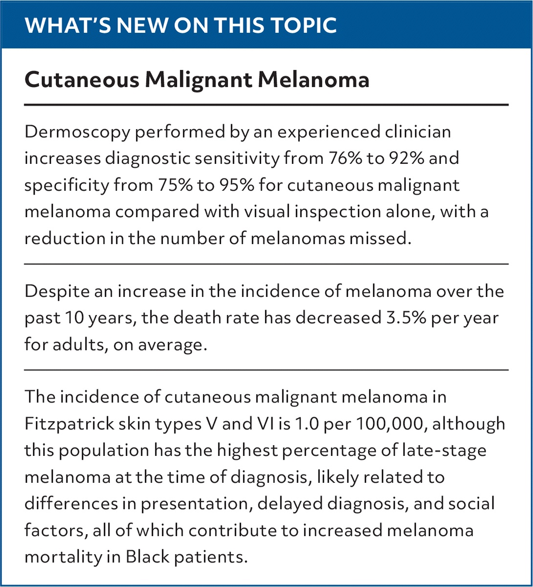
| Dermoscopy performed by an experienced clinician increases diagnostic sensitivity from 76% to 92% and specificity from 75% to 95% for cutaneous malignant melanoma compared with visual inspection alone, with a reduction in the number of melanomas missed. |
| Despite an increase in the incidence of melanoma over the past 10 years, the death rate has decreased 3.5% per year for adults, on average. |
| The incidence of cutaneous malignant melanoma in Fitzpatrick skin types V and VI is 1.0 per 100,000, although this population has the highest percentage of late-stage melanoma at the time of diagnosis, likely related to differences in presentation, delayed diagnosis, and social factors, all of which contribute to increased melanoma mortality in Black patients. |
SORT: KEY RECOMMENDATIONS FOR PRACTICE
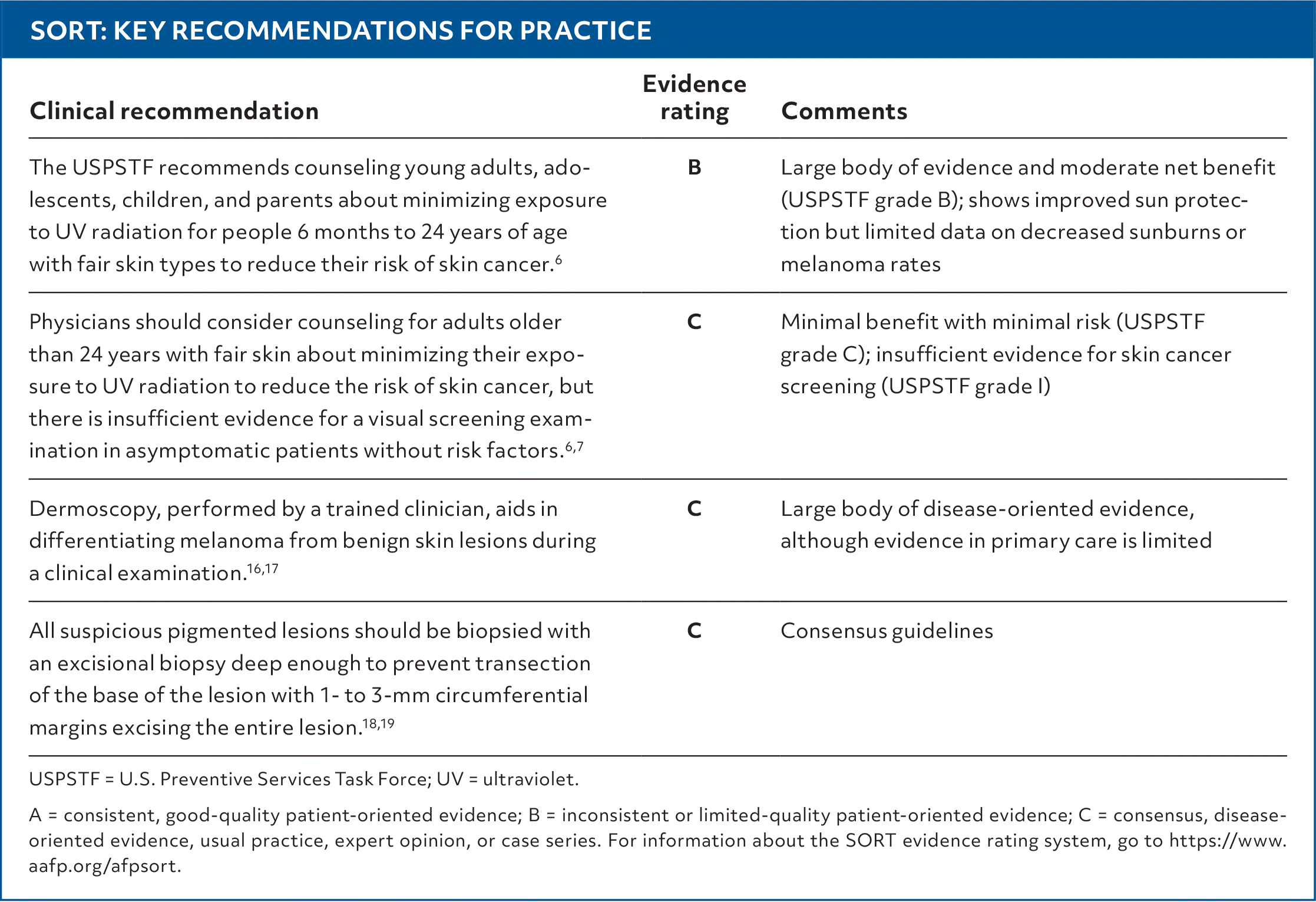
USPSTF = U.S. Preventive Services Task Force; UV = ultraviolet.
A = consistent, good-quality patient-oriented evidence; B = inconsistent or limited-quality patient-oriented evidence; C = consensus, disease-oriented evidence, usual practice, expert opinion, or case series. For information about the SORT evidence rating system, go to https://www.aafp.org/afpsort.
Read the full article
Get immediate access, anytime, anywhere.
Choose a single article, issue, or full-access subscription.
Earn up to 14 CME credits per issue.
