

Acid-Base Interpretation: A Practical Approach

Masahiro J. Morikawa, MD, MPH
Prakash R. Ganesh, MD, MPH

American Family Physician. 2025;111(2):148-155.

Author disclosure: No relevant financial relationships.

This clinical content conforms to AAFP criteria for CME.

Acid-base disorders are prevalent in critically ill patients, and a systematic approach is essential for evaluation. The first step is to determine the primary process based on a patient's pH, partial pressure of carbon dioxide, and bicarbonate measurements. After this is complete, the next step is to evaluate for respiratory or metabolic compensation. Deviations from expected compensation may indicate additional acid-base processes. For metabolic acidosis, anion gap calculation distinguishes between anion gap metabolic acidosis and non–anion gap metabolic acidosis. The evaluation for anion gap metabolic acidosis includes calculating the osmolal gap and conducting a gap-gap analysis. Evaluating non–anion gap metabolic acidosis involves urine anion gap calculation. These analyses identify potential etiologies and additional acid-base disturbances. Metabolic alkalosis assessment begins with measuring urine chloride levels to determine whether the process is a result of chloride depletion. Respiratory acidosis, caused by hypoventilation, often results from chronic lung disease or neuromuscular dysfunction. Respiratory alkalosis, due to hyperventilation, is common in sepsis, chronic liver disease, and acute pulmonary embolism.

Acid-base disorders are common in patients who are critically ill.1 A systematic approach to evaluating these disorders provides vital diagnostic information and guides timely treatment.25 Acidosis leads to acidemia (pH less than 7.38)6 and is typically due to one of three factors: (1) accumulation of acids (eg, salicylic acid, keto acids, lactic acid) or loss of alkali (as in diarrhea or proximal renal tubular acidosis); (2) decreased renal excretion of acid (as in kidney failure or distal renal tubular acidosis); or (3) accumulation of carbon dioxide in the body. Alkalosis is a process that leads to alkalemia (pH greater than 7.42) and is primarily caused by excessive carbon dioxide exhalation (respiratory alkalosis), the accumulation of alkali (sodium bicarbonate [HCO3] administration), or loss of acid (as with gastrointestinal loss from vomiting and diarrhea).7

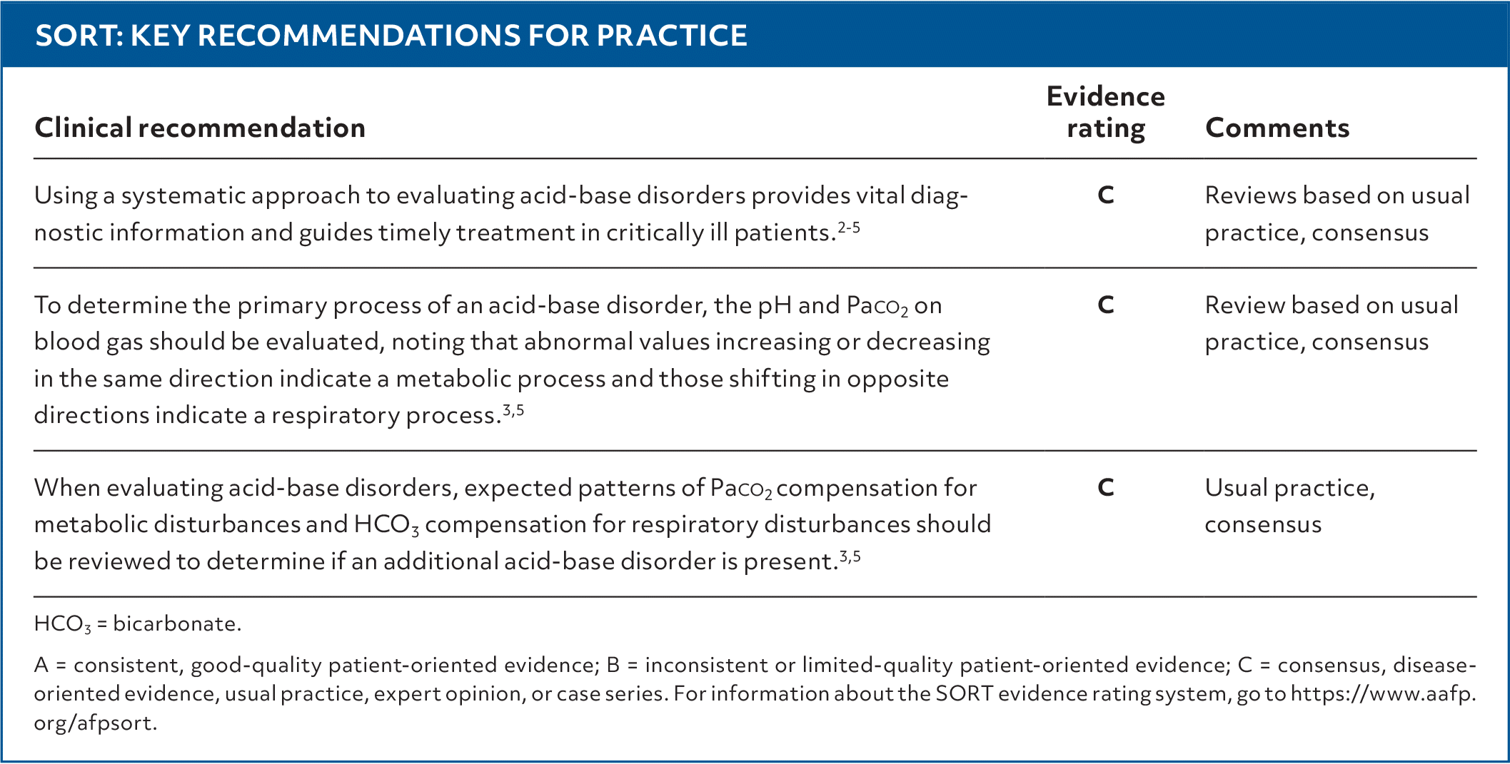
SORT: KEY RECOMMENDATIONS FOR PRACTICE
Clinical recommendationEvidence ratingComments
Using a systematic approach to evaluating acid-base disorders provides vital diagnostic information and guides timely treatment in critically ill patients.25 CReviews based on usual practice, consensus
To determine the primary process of an acid-base disorder, the pH and Paco2 on blood gas should be evaluated, noting that abnormal values increasing or decreasing in the same direction indicate a metabolic process and those shifting in opposite directions indicate a respiratory process.3,5 CReview based on usual practice, consensus
When evaluating acid-base disorders, expected patterns of Paco2 compensation for metabolic disturbances and HCO3 compensation for respiratory disturbances should be reviewed to determine if an additional acid-base disorder is present.3,5 CUsual practice, consensus

HCO3 = bicarbonate.

A = consistent, good-quality patient-oriented evidence; B = inconsistent or limited-quality patient-oriented evidence; C = consensus, disease-oriented evidence, usual practice, expert opinion, or case series. For information about the SORT evidence rating system, go to https://www.aafp.org/afpsort.

APPROACH

The physiologic approach to acid-base analysis is based on the concept of the carbonic acid-HCO3 buffer system as a center of homeostatic control of blood pH.24,8,9 In addition to assessment of vital signs and fluid status, blood gas measurement and basic chemistry panel are required. Venous blood gas often is measured initially instead of arterial blood gas because it is easier to obtain and may reveal metabolic changes earlier in cases of circulatory insufficiency.10 An arterial blood gas measurement is preferred when assessing a patient's oxygenation status.

MASAHIRO J. MORIKAWA, MD, MPH, is the B. Lewis Barnett Jr Professor of Family Medicine at the University of Virginia, Charlottesville.

PRAKASH R. GANESH, MD, MPH, is an assistant professor in the Department of Family Medicine & Community Health at the University Hospitals Cleveland Medical Center, and at the Center for Community Health Integration, Case Western Reserve University School of Medicine, and Neighborhood Family Practice, Ohio

Address correspondence to Masahiro J. Morikawa, MD, MPH, at mm7jc@virginia.edu.

Author disclosure: No relevant financial relationships.

  1. 1.Djamali A, Singh T, Melamed ML, et al. Metabolic acidosis 1 year following kidney transplantation and subsequent cardiovascular events and mortality: an observational cohort study. Am J Kidney Dis. 2019;73(4):476-485.
  2. 2.Berend K, de Vries APJ, Gans ROB. Physiological approach to assessment of acid-base disturbances. N Engl J Med. 2014;371(15):1434-1445.
  3. 3.Achanti A, Szerlip HM. Acid-base disorders in the critically ill patient. Clin J Am Soc Nephrol. 2023;18(1):102-112.
  4. 4.Whittier WL, Rutecki GW. Primer on clinical acid-base problem solving. Dis Mon. 2004;50(3):122-162.
  5. 5.Palmer BF. Approach to fluid and electrolyte disorders and acid-base problems. Prim Care. 2008;35(2):195-213.
  6. 6.Berend K. Acid-base pathophysiology after 130 years: confusing, irrational and controversial. J Nephrol. 2013;26(2):254-265.
  7. 7.Hamm LL, Nakhoul N, Hering-Smith KS. Acid-base homeostasis. Clin J Am Soc Nephrol. 2015;10(12):2232-2242.
  8. 8.Gomez H, Kellum JA. Understanding acid base disorders. Crit Care Clin. 2015;31(4):849-860.
  9. 9.Haber RJ. A practical approach to acid-base disorders. West J Med. 1991;155(2):146-151.
  10. 10.Gauthier PM, Szerlip HM. Metabolic acidosis in the intensive care unit. Crit Care Clin. 2002;18(2):289-308.
  11. 11.Kamel KS, Halperin ML. Fluid, Electrolyte, and Acid-Base Physiology. 5th ed. Elsevier; 2016.
  12. 12.Casaletto JJ. Differential diagnosis of metabolic acidosis. Emerg Med Clin North Am. 2005;23(3):771-787.
  13. 13.Kraut JA, Madias NE. Serum anion gap: its uses and limitations in clinical medicine. Clin J Am Soc Nephrol. 2007;2(1):162-174.
  14. 14.Chabali R. Diagnostic use of anion and osmolal gaps in pediatric emergency medicine. Pediatr Emerg Care. 1997;13(3):204-210.
  15. 15.Martin L. Primary and mixed acid-base disorders. All You Really Need to Know to Interpret Arterial Blood Gases. 2nd ed. Lippincott Williams & Wilkins; 1999: 137.
  16. 16.Rastegar A. Use of the DeltaAG/DeltaHCO3− ratio in the diagnosis of mixed acid-base disorders. J Am Soc Nephrol. 2007;18(9):2429-2431.
  17. 17.Paulson WD, Gadallah MF. Diagnosis of mixed acid-base disorders in diabetic ketoacidosis. Am J Med Sci. 1993;306(5):295-300.
  18. 18.Gennari FJ. Pathophysiology of metabolic alkalosis: a new classification based on the centrality of stimulated collecting duct ion transport. Am J Kidney Dis. 2011;58(4):626-636.
  19. 19.Reddy P, Mooradian AD. Clinical utility of anion gap in deciphering acid-base disorders. Int J Clin Pract. 2009;63(10):1516-1525.
  20. 20.Judge BS. Differentiating the causes of metabolic acidosis in the poisoned patient. Clin Lab Med. 2006;26(1):31-48.
  21. 21.Kraut JA, Madias NE. Differential diagnosis of nongap metabolic acidosis: value of a systematic approach. Clin J Am Soc Nephrol. 2012;7(4):671-679.
  22. 22.Yunos NM, Kim IB, Bellomo R, et al. The biochemical effects of restricting chloride-rich fluids in intensive care. Crit Care Med. 2011;39(11):2419-2424.
  23. 23.Schreiber MA. The use of normal saline for resuscitation in trauma. J Trauma. 2011;70(5 suppl):S13-S14.
  24. 24.Lafrance JP, Leblanc M. Metabolic, electrolytes, and nutritional concerns in critical illness. Crit Care Clin. 2005;21(2):305-327.
  25. 25.Rodríguez Soriano J. Renal tubular acidosis: the clinical entity. J Am Soc Nephrol. 2002;13(8):2160-2170.
  26. 26.Soleimani M, Rastegar A. Pathophysiology of renal tubular acidosis: core curriculum 2016. Am J Kidney Dis. 2016;68(3):488-498.
  27. 27.Giglio S, Montini G, Trepiccione F, et al. Distal renal tubular acidosis: a systematic approach from diagnosis to treatment. J Nephrol. 2021;34(6):2073-2083.
  28. 28.Batlle DC, Hizon M, Cohen E, et al. The use of the urinary anion gap in the diagnosis of hyperchloremic metabolic acidosis. N Engl J Med. 1988;318(10):594-599.
  29. 29.Luke RG, Galla JH. It is chloride depletion alkalosis, not contraction alkalosis. J Am Soc Nephrol. 2012;23(2):204-207.
  30. 30.Emmett M. Metabolic alkalosis. A brief pathophysiologic review. Clin J Am Soc Nephrol. 2020;15(12):1848-1856.
  31. 31.Fernandes A, Falcão L, Raimundo M. A severe case of hypokalemic metabolic alkalosis: a quiz. Am J Kidney Dis. 2023;81(4):A13-A16.
  32. 32.Do C, Vasquez PC, Soleimani M. Metabolic alkalosis pathogenesis, diagnosis, and treatment: core curriculum 2022. Am J Kidney Dis. 2022;80(4):536-551.

Copyright © 2026 by the American Academy of Family Physicians.

This content is owned by the AAFP. A person viewing it online may make one printout of the material and may use that printout only for his or her personal, non-commercial reference. This material may not otherwise be downloaded, copied, printed, stored, transmitted or reproduced in any medium, whether now known or later invented, except as authorized in writing by the AAFP. See permissions for copyright questions and/or permission requests.
