

Developmental dysplasia of the hip (DDH) is the most common joint condition in infants, encompassing a complex spectrum of pathologic states that can result in hip instability and dislocation. The most significant risk factors for DDH are breech positioning in the third trimester and family history of hip dysplasia. The condition is more common in females. Diagnosis is based on age-specific physical examination maneuvers and imaging studies. The Ortolani and Barlow maneuvers are the recommended physical examination techniques used to screen infants up to 3 months of age. Ultrasonography is the preferred imaging modality to evaluate younger infants, whereas plain radiography is preferred after 4 months of age. If DDH is identified, abduction bracing is the first-line treatment in infants younger than 6 months. Operative management is reserved for infants older than 6 months or if abduction bracing fails. Early diagnosis may prevent the need for invasive surgical procedures and reduce the risk of early degenerative changes of the hip in adulthood.
Developmental dysplasia of the hip (DDH), the most common joint condition in infants, encompasses a complex spectrum of hip joint abnormalities. This ranges from mild instability (static or dynamic), to subluxation, to frank dislocation caused by capsular laxity and mechanical instability. DDH can also lead to femoral head incongruency (ie, the femoral head no longer fits properly within the acetabular socket) as a late sequela of dislocation.
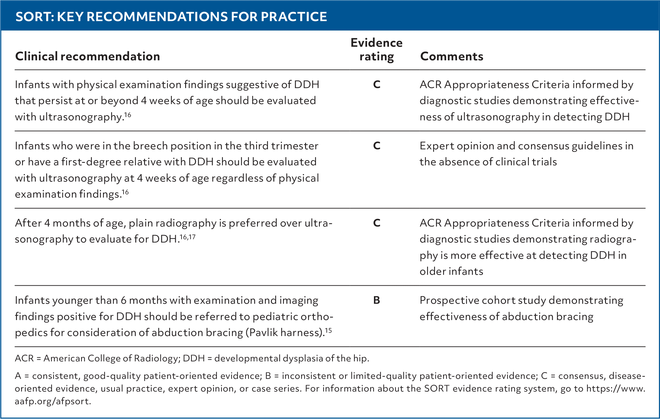
SORT: KEY RECOMMENDATIONS FOR PRACTICE
| Clinical recommendation | Evidence rating | Comments |
|---|---|---|
| Infants with physical examination findings suggestive of DDH that persist at or beyond 4 weeks of age should be evaluated with ultrasonography.16 | C | ACR Appropriateness Criteria informed by diagnostic studies demonstrating effectiveness of ultrasonography in detecting DDH |
| Infants who were in the breech position in the third trimester or have a first-degree relative with DDH should be evaluated with ultrasonography at 4 weeks of age regardless of physical examination findings.16 | C | Expert opinion and consensus guidelines in the absence of clinical trials |
| After 4 months of age, plain radiography is preferred over ultrasonography to evaluate for DDH.16,17 | C | ACR Appropriateness Criteria informed by diagnostic studies demonstrating radiography is more effective at detecting DDH in older infants |
| Infants younger than 6 months with examination and imaging findings positive for DDH should be referred to pediatric orthopedics for consideration of abduction bracing (Pavlik harness).15 | B | Prospective cohort study demonstrating effectiveness of abduction bracing |
ACR = American College of Radiology; DDH = developmental dysplasia of the hip.
A = consistent, good-quality patient-oriented evidence; B = inconsistent or limited-quality patient-oriented evidence; C = consensus, disease-oriented evidence, usual practice, expert opinion, or case series. For information about the SORT evidence rating system, go to https://www.aafp.org/afpsort.
DDH is generally recognized as a condition of infancy, primarily attributed to in utero positioning and intrinsic risk factors. There is no consensus as to which measurable findings, and at what ages, constitute normal variation vs pathology.1 However, if the hip is not evaluated in infancy, DDH may remain undiagnosed until the infant becomes mobile or into childhood or adolescence, when conservative management may no longer be effective. Even when symptoms of hip dysplasia do not develop until later in childhood or adolescence, DDH was present in infancy.
Read the full article
Get immediate access, anytime, anywhere.
Choose a single article, issue, or full-access subscription.
Earn up to 14 CME credits per issue.
