

Vertebral compression fracture is a common complication of osteoporosis. It is often triggered by ordinary behaviors such as turning in bed, coughing, and sneezing, but traumatic or metastatic etiologies are also possible. Although patients with vertebral compression fractures are often asymptomatic, they can present with back pain that worsens with postural movement and the Valsalva maneuver, potentially impairing function. Long term, these fractures can cause kyphosis, decreased vertebral height, muscle atrophy, and further bone mineral density loss. Anteroposterior and lateral projection radiography of the spine should be the initial imaging modality, and magnetic resonance imaging can be used to confirm suspicious but radiography-negative cases. Conservative measures are the mainstay of treatment, with physical rehabilitation and pharmacotherapy for pain relief. In addition to nonsteroidal anti-inflammatory drugs and acetaminophen, several anti-osteoporotic medications can improve pain after fracture. Bracing and nerve root blocks have very limited evidence of short-term benefit. Surgical intervention with vertebroplasty or kyphoplasty can be considered when pain persists for 6 weeks despite conservative intervention. Prevention of low bone mineral density is critical for avoiding vertebral compression fractures.
Vertebral compression fracture is the most common complication of osteoporosis, accounting for 1.4 million cases worldwide and approximately 700,000 cases annually in the United States.1–3 Bone mineral density loss, including osteopenia and osteoporosis, is a major risk factor for compression fracture.1,2 Vertebral compression fractures are categorized as fragility fractures and are diagnostic for osteoporosis.3
SORT: KEY RECOMMENDATIONS FOR PRACTICE
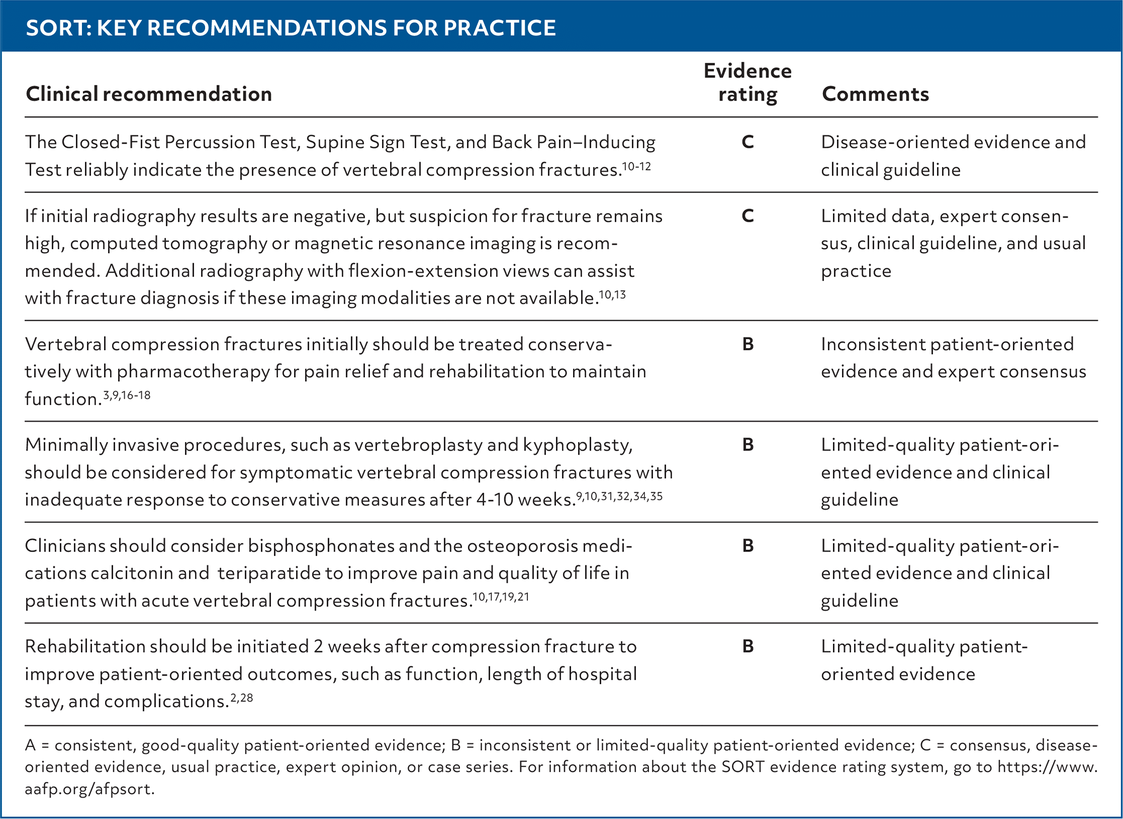
| Clinical recommendation | Evidence rating | Comments |
|---|---|---|
| The Closed-Fist Percussion Test, Supine Sign Test, and Back Pain–Inducing Test reliably indicate the presence of vertebral compression fractures.10–12 | C | Disease-oriented evidence and clinical guideline |
| If initial radiography results are negative, but suspicion for fracture remains high, computed tomography or magnetic resonance imaging is recommended. Additional radiography with flexion-extension views can assist with fracture diagnosis if these imaging modalities are not available.10,13 | C | Limited data, expert consensus, clinical guideline, and usual practice |
| Vertebral compression fractures initially should be treated conservatively with pharmacotherapy for pain relief and rehabilitation to maintain function.3,9,16–18 | B | Inconsistent patient-oriented evidence and expert consensus |
| Minimally invasive procedures, such as vertebroplasty and kyphoplasty, should be considered for symptomatic vertebral compression fractures with inadequate response to conservative measures after 4–10 weeks.9,10,31,32,34,35 | B | Limited-quality patient-oriented evidence and clinical guideline |
| Clinicians should consider bisphosphonates and the osteoporosis medications calcitonin and teriparatide to improve pain and quality of life in patients with acute vertebral compression fractures.10,17,19,21 | B | Limited-quality patient-oriented evidence and clinical guideline |
| Rehabilitation should be initiated 2 weeks after compression fracture to improve patient-oriented outcomes, such as function, length of hospital stay, and complications.2,28 | B | Limited-quality patient-oriented evidence |
A = consistent, good-quality patient-oriented evidence; B = inconsistent or limited-quality patient-oriented evidence; C = consensus, disease-oriented evidence, usual practice, expert opinion, or case series. For information about the SORT evidence rating system, go to https://www.aafp.org/afpsort.
Read the full article
Get immediate access, anytime, anywhere.
Choose a single article, issue, or full-access subscription.
Earn up to 14 CME credits per issue.
