

A 4-day-old female infant was brought to the clinic for evaluation of a blue nodule near her left eye. The lesion had been noted at birth and had gradually increased in size. There was no drainage or bleeding. The infant was born at 40 weeks of gestation by uncomplicated vaginal delivery following an uncomplicated pregnancy.
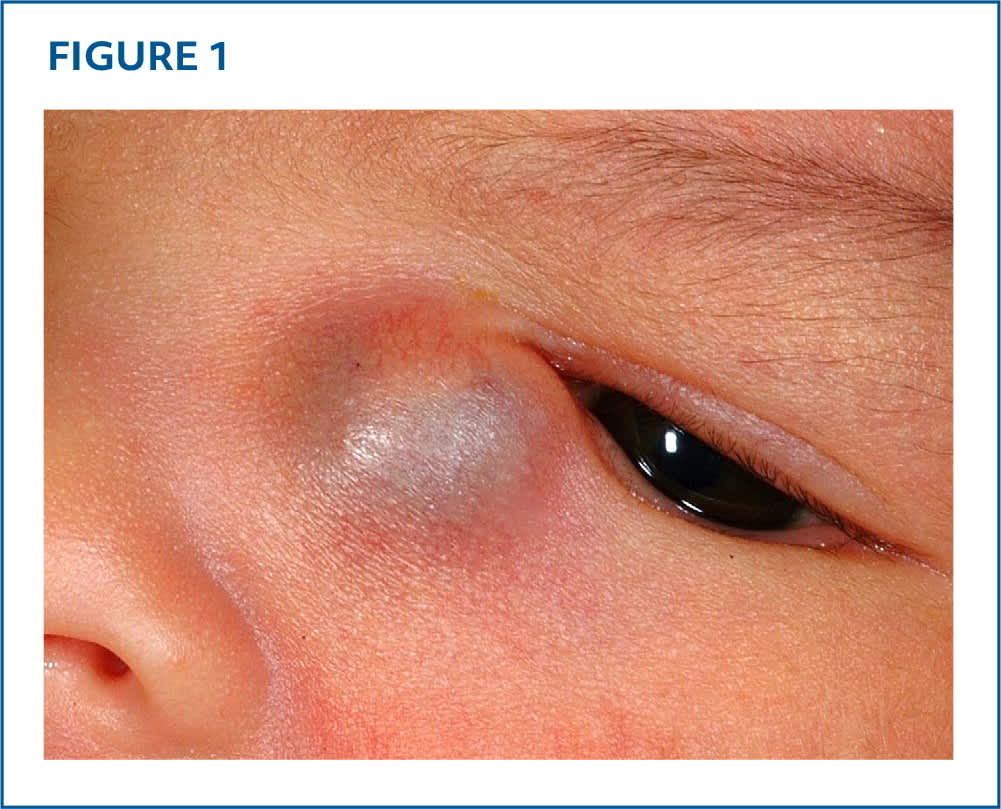
Physical examination revealed a cystic, minimally compressible, blue nodule on the left inferior medial canthus (Figure 1). The nodule exhibited slight transillumination and was 12 × 11 mm. Some associated tearing was noted, but no visual axis obstruction was present.
FIGURE 1
Question
Based on the patient's history and physical examination, which one of the following is the most likely diagnosis?
- A. Blue nevus.
- B. Cephalocele.
- C. Dacryocystocele.
- D. Infantile hemangioma.
- E. Neuroblastoma.
DISCUSSION
The answer is C: dacryocystocele, a cyst caused by congenital nasolacrimal duct obstruction. It presents as a blue to pink cystic mass in the medial canthal region and may be associated with epiphora and dacryocystitis. Dacryocystocele can mimic other conditions, including deep infantile hemangioma, dermoid cyst, cephalocele, and glioma.1 Conservative monitoring or manual massage are suitable for uncomplicated cases, but surgical intervention may be necessary for larger lesions or those associated with inflammation or infection.2
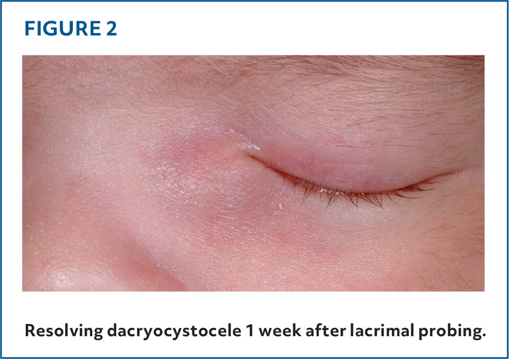
Ophthalmologic evaluation and computed tomography confirmed dacryocystocele in this patient. Lacrimal probing was performed, and 1 week later, the dacryocystocele was almost fully resolved (Figure 2).
FIGURE 2. Resolving dacryocystocele 1 week after lacrimal probing.
Blue nevus is a solitary, well-demarcated, blue or blue-black papule or nodule caused by dermal melanocyte proliferation. It typically occurs on the face or extremities. Lesions are usually acquired but may be congenital. Nevi are typically firm, noncystic, asymptomatic, and static.3,4 Transillumination is not expected in a blue nevus.
A cephalocele is a birth defect characterized by the protrusion of the meninges with or without brain tissue through a congenital skull defect. Cephaloceles are categorized according to their content and skull defect location. They typically present as midline facial lesions (frontonasal cephalocele) and are less common in the medial canthus area. A cephalocele is often compressible, can pulsate or enlarge with crying or straining, and may be associated with hydrocephalus, seizures, and other congenital anomalies.5
Infantile hemangioma is a common benign tumor that develops from vascular endothelial cells. It may present as a bright red lesion (superficial type), blue nodular lesion (deep type), or a combination.6 Hemangiomas are usually not present at birth but appear in the first few weeks of life. They are most common in preterm female infants and are associated with superficial telangiectasias. Hemangiomas are soft and compressible (but not cystic), and typically do not transilluminate or feature epiphora.
Neuroblastoma is a malignant tumor of neuroblasts that usually occurs in the adrenal glands and can spread to multiple organs, including the skin, where it can present as small, solid blue papules or nodules with a “blueberry muffin” appearance.7 These lesions are typically small, multiple, and disseminated and usually do not transilluminate.
SUMMARY TABLE
| Condition | Characteristics |
|---|---|
| Blue nevus | Solitary, well-demarcated, blue or blue-black papule or nodule; acquired (rarely congenital), solitary, noncystic, asymptomatic, and static |
| Cephalocele | Birth defect; most commonly manifests as a midline facial lesion; compressible, pulsates or enlarges with crying or straining, and may be associated with other neurologic findings |
| Dacryocystocele | Congenital lesion, typically distributed in the medial canthus; blue to pink, cystic, and partially compressible; may enlarge, transilluminate, and be associated with epiphora, inflammation (dacrocystitis), and nasal obstruction |
| Infantile hemangioma | Typically appears in the first weeks of life; bright red to blue, soft, compressible (but not cystic); typically does not transilluminate; associated with superficial telangiectasias |
| Neuroblastoma | Malignant tumor of neuroblasts; metastatic disease may present as multiple disseminated, small, solid blue papules or nodules; typically does not transilluminate |
Read the full article
Get immediate access, anytime, anywhere.
Choose a single article, issue, or full-access subscription.
Earn up to 14 CME credits per issue.
