

A 14-year old boy presented with a 1-year history of pain and irritation in the nail area of both great toes. He was prescribed efinaconazole 10% topical solution (Jublia). The patient returned 2 months later with greenish discoloration of the affected toenails. Bacterial culture grew an aerobic, gram-negative organism. Gentamicin 0.3% eye drops were prescribed to be applied to the nail edges three times per day. The patient returned 1 month later without improvement.
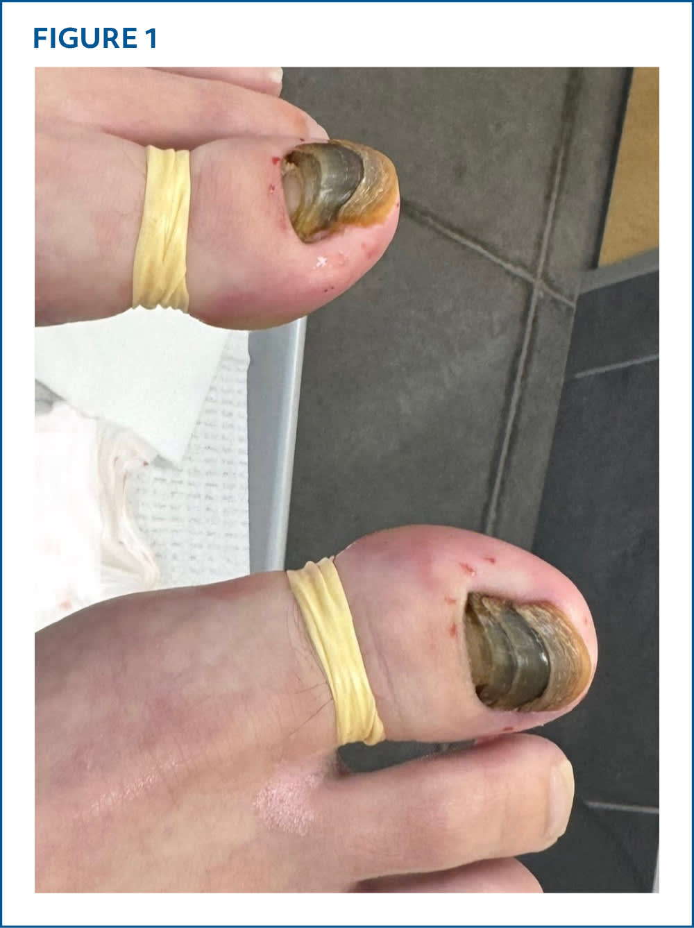
Physical examination revealed yellow-green discolored nails with onycholysis and subungual debris on both great toes. The nails had layered growth with a stacked appearance (Figure 1).
FIGURE 1
QUESTION
Based on the patient's history and physical examination, which one of the following is the most likely diagnosis?
- A. Green nail syndrome.
- B. Onychocryptosis (ingrown toenail).
- C. Onychomadesis.
- D. Refractory onychomycosis.
- E. Retronychia.
DISCUSSION
The answer is E: retronychia, a pathologic pattern of nail growth in which the nail plate embeds retrograde into the proximal nail fold.1,2 This aberrant ingrowth leads to a characteristic stacking of successive generations of nail plates underneath the proximal nail fold.2 Retronychia is a clinical diagnosis and should be suspected in the setting of ongoing paronychia, granulation tissue emerging from under the nail fold, thickening at the proximal nail plate, xanthonychia, and atypical, layered nail plate growth.3
Retronychia is a rare condition most commonly caused by repetitive trauma. The great toe is usually affected, likely attributable to its vulnerability to trauma through everyday activities. Risk factors include wearing poorly fitting shoes or high heels and performing exertional physical activity. Ischemic etiologies, such as compartment syndrome, are much less common.2 The definitive management of retronychia is complete nail avulsion.
Green nail syndrome is a bacterial infection of the nails, typically due to Pseudomonas aeruginosa, that leads to green discoloration, erythema, edema, and pain. It is associated with persistent exposure of the nails to wet environments and trauma.4 A pseudomonal nail infection may be a complication of retronychia.5
Onychocryptosis (ingrown toenail) occurs when the nail plate embeds into the adjacent periungual soft tissues. Like retronychia, it may present with erythema, edema, and pain. The great toe is most commonly affected. Trauma, inadequate foot hygiene, improperly fitting footwear, and hyperhidrosis are risk factors.6
Onychomadesis describes a pathologic process in which growth from the nail matrix is completely halted, which leads to deep, horizontal depressions across the nail plate that may cause it to break from the base. Onychomadesis may arise as a sequela of viral infection (eg, hand-foot-and-mouth disease due to coxsackievirus), other systemic illnesses, trauma, and medications.7
Refractory onychomycosis is a persistent fungal infection of the nails that is resistant to conventional first- and second-line treatments. First-line antifungal agents include oral terbinafine and itraconazole. Diagnosis may require a full diagnostic workup, including fungal culture, microscopy, and polymerase chain reaction testing to identify the causative pathogen.8
SUMMARY TABLE
| Condition | Characteristics |
|---|---|
| Green nail syndrome | Bacterial infection of nails, typically due to Pseudomonas aeruginosa; green discoloration of nail plate; erythema, edema, and pain |
| Onychocryptosis (ingrown toenail) | Nail plate embeds into adjacent periungual tissue; erythema, edema, and pain |
| Onychomadesis | Arrested growth from the nail matrix; nail plate may break from the base; deep, horizontal depressions in nail plate |
| Refractory onychomycosis | Fungal infection of the nails that does not respond to first- and second-line treatments; may require a full diagnostic workup |
| Retronychia | Nail plate embeds retrograde into the proximal nail fold with stacking of successive generations of nail plates; paronychia, xanthonychia, and granulation tissue formation |
Read the full article
Get immediate access, anytime, anywhere.
Choose a single article, issue, or full-access subscription.
Earn up to 14 CME credits per issue.
