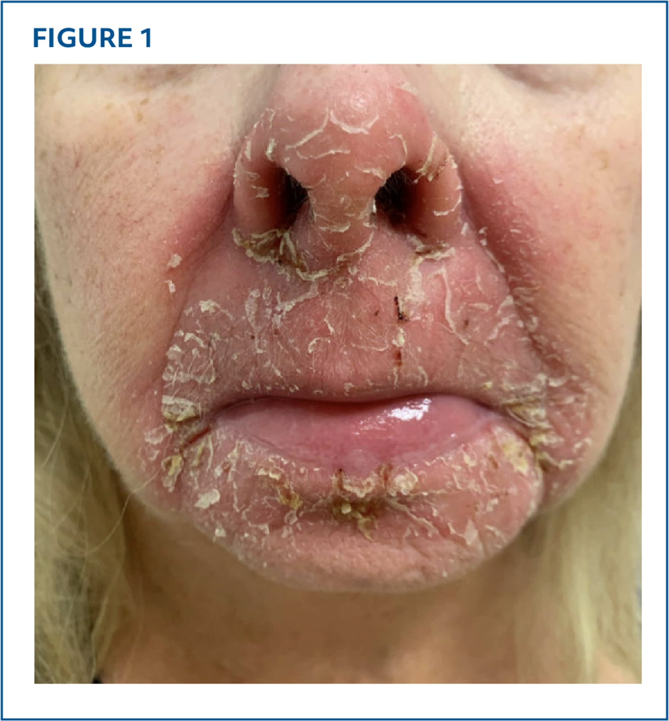
A 68-year-old woman presented for evaluation of a pruritic, burning rash around her mouth and nose that first developed 4 years earlier. She had severe flare-ups approximately every 2 weeks but no worsening of symptoms following exposure to sunlight or consumption of alcohol. The rash has been treated with oral antibiotics, and intramuscular and topical corticosteroids with only temporary symptom relief. The patient had applied an over-the-counter mentholated topical ointment around her nose and mouth approximately twice a month for more than 50 years but used no other products on her face. She had no known allergies to cosmetics, personal care products, or synthetic materials.
Physical examination revealed well-defined, pink, scaly plaques with fissures on the central and lower face (Figure 1).
FIGURE 1
QUESTION
Based on the patient's history and physical examination, which one of the following is the most likely diagnosis?
- A. Allergic contact dermatitis.
- B. Perioral dermatitis.
- C. Rosacea.
- D. Seborrheic dermatitis.
- E. Tinea faciei.
DISCUSSION
The answer is A: allergic contact dermatitis. The chronicity, well-demarcated eczematous plaques, and fissuring support the diagnosis of allergic contact dermatitis. This was confirmed by a patch test with a positive reaction to turpentine oil.
Allergic contact dermatitis affects approximately 20% of the general population.1 The condition is more common in individuals with occupational exposure to sensitizing agents, including health care professionals, hairdressers, cleaners, industrial workers, and machinists.2 Allergic contact dermatitis is a delayed (type IV) hypersensitivity reaction mediated by allergen-specific T lymphocytes. Sensitization occurs when haptens penetrate the epidermis, bind to proteins, and form complete antigens that are presented by Langerhans cells to T cells.3 Repeated or prolonged exposure may be required before sensitization occurs, as in this case.
Allergic contact dermatitis most commonly presents as pruritic, scaly, erythematous papules and plaques. It may appear vesicular with crusting or bullae. Lichenification is common with chronic allergic contact dermatitis due to repeated inflammation and scratching. Symptoms of acute allergic contact dermatitis typically develop 24 to 72 hours after allergen exposure and reach peak intensity between 48 and 72 hours.3
Epicutaneous patch testing is the standard diagnostic tool for allergic contact dermatitis.4 Primary treatment is avoidance of the allergen. If contact occurs, symptoms should be treated with topical medications such as corticosteroids, calcineurin inhibitors, or phosphodiesterase-4 inhibitors. Systemic therapy such as an interleukin-4 or interleukin-13 inhibitor may be necessary if topical therapy is ineffective.
Perioral dermatitis presents as erythematous papules and pustules centered around the mouth, nose, and sometimes the eyelids. Use of topical corticosteroids often triggers or exacerbates rather than improves symptoms.5
Rosacea also causes persistent centrofacial erythema but often with inflammatory papules or pustules, telangiectasias, and ocular symptoms.6 Common triggers of rosacea symptoms include sunlight, stress, alcohol, and spicy foods.
Seborrheic dermatitis is a chronic papulosquamous condition that develops in sebaceous-rich areas and is associated with Malassezia species. Typical findings include erythematous plaques with greasy, yellow scale often involving the scalp, eyebrows, ears, nasolabial folds, and chest.7
Tinea faciei is caused by a dermatophyte fungal infection and typically presents as erythematous, annular plaques with central clearing and an active, scaly border. Dermatophyte infection usually worsens with topical corticosteroid use due to tinea incognito.8
Read the full article
Get immediate access, anytime, anywhere.
Choose a single article, issue, or full-access subscription.
Earn up to 14 CME credits per issue.