

A 71-year-old woman presented for routine follow-up after a skin lesion was noted on her right cheek; it had appeared 6 months prior. The patient said it began as acne, bled when scratched, and then crusted. After healing, the lesion persisted and was nonpruritic. The patient had no personal or family history of skin cancer. She had a history of colon cancer in her late 30s that was treated with chemotherapy and radiation.
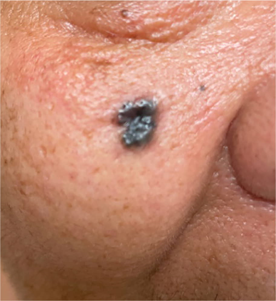
Physical examination revealed a uniformly brown papule with rolled borders on her right cheek measuring 7 × 6 mm (Figure 1). A biopsy was performed.
FIGURE 1
QUESTION
Based on the patient's history and physical examination, which one of the following is the most likely diagnosis?
- A. Basal cell carcinoma.
- B. Lentigo.
- C. Melanoma.
- D. Nevus.
- E. Squamous cell carcinoma.
DISCUSSION
The answer is A: basal cell carcinoma, which is the most common type of skin cancer and originates in the basal cell layer of the skin.1 It is more common in patients with lighter skin tones and is usually found on sun-exposed areas such as the face and neck. In patients with darker skin tones, basal cell carcinoma often presents as pearly black lesions. In patients with lighter skin tones, it typically presents as pearly papules with central ulceration and telangiectasias.2
Nodular, superficial, and morphea forms are the most common subtypes of basal cell carcinoma. The nodular subtype was diagnosed in this patient by histologic review of the biopsy specimen. The nodular subtype represents more than one-half of all diagnosed basal cell carcinomas. Up to 90% of lesions in the nodular subtype are located on the head.3
Lentigo skin lesions are typically small, flat, tan-brown macules with asymmetrical, irregular borders and are greater than 1 cm in diameter. They are often referred to as liver spots. Lentigines are found on chronically sun-damaged skin in exposed areas such as the hands, arms, and face. They can be malignant.4
Melanoma is an aggressive form of skin cancer caused by melanocytes. Melanoma is more common in those with a family history of the condition. Other risk factors include lighter skin tones and excessive sun exposure. The ABCD mnemonic is used to describe the common characteristics of a melanoma5:
- Asymmetry
- Borders (irregular)
- Color (variable or changing)
- Diameter (greater than 6 mm)
Nevi are small macules or papules characterized by their uniformity in shape and size and their brown or black color. They are found on any part of the body. They are generally benign, but a lesion greater than 6 mm in diameter should be considered atypical with a higher risk of melanoma. The presence of numerous nevi (more than 50) is also a risk factor for melanoma.6
Squamous cell carcinoma presents as rough, scaly, firm papules on an erythematous base in patients with lighter skin tones. They are more common on sun-exposed areas, such as the back of the hands and forearms. In patients with darker skin tones, lesions may present as nonhealing sores with bleeding and crusting.2 Squamous cell carcinoma is more common in patients older than 50 years.7
Read the full article
Get immediate access, anytime, anywhere.
Choose a single article, issue, or full-access subscription.
Earn up to 14 CME credits per issue.
